Products Found:
13
Showing Results For:
Slides, adhesive
Slides, adhesive
Single Product
Single Product
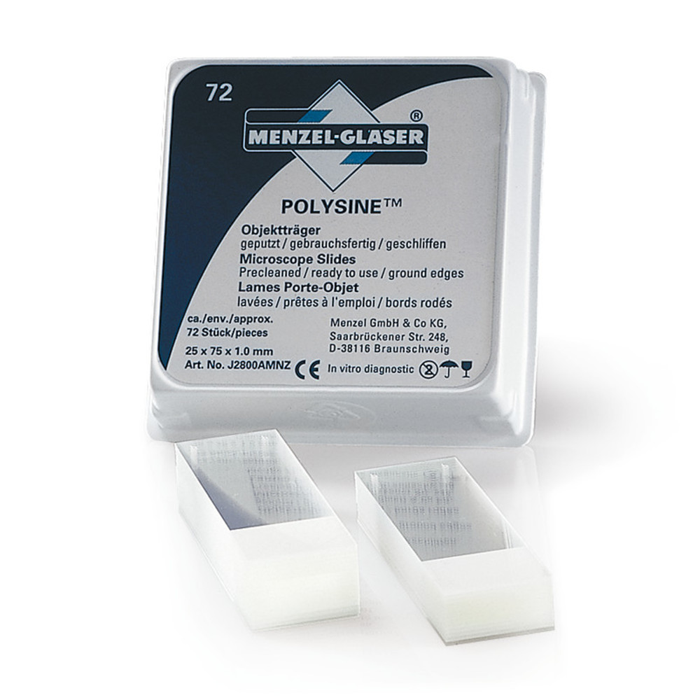
Adhesion slides Superfrost ® Plus / AC100060765
MF Part:
H867.1
MOQ:
1
£ 25.61
In Stock
more details
Multiple Variants

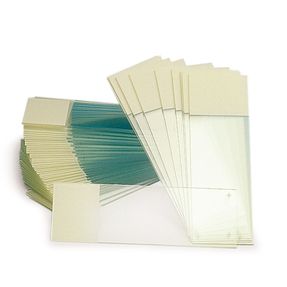
Adhesion slides ROTI ® Bond, 5 unit(s) / AC100042070
MF Part:
CL20.1
MOQ:
1
£ 11.12
In Stock
more details
Multiple Variants

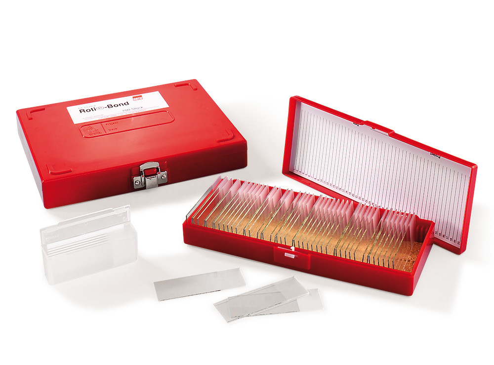
Adhesion slides ROTI ® Bond, 50 unit(s) / AC100040517
MF Part:
CL20.2
MOQ:
1
£ 87.62
In Stock
more details
Multiple Variants
Adhesion slides ROTI ® Bond, 100 unit(s) / AC100038758
MF Part:
CL20.3
MOQ:
1
£ 136.76
In Stock
more details
Single Product
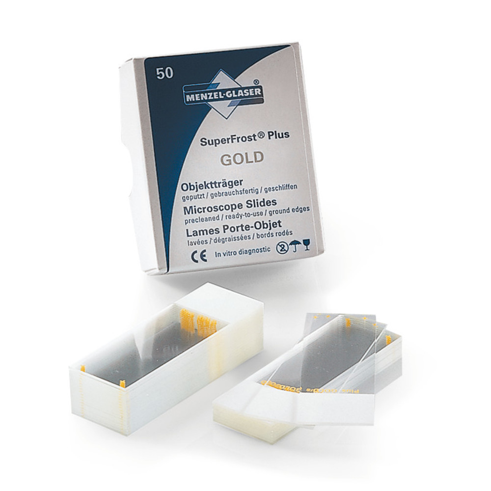
Adhesion slides Superfrost ® Plus Gold / AC100045094
MF Part:
ET09.2
MOQ:
1
£ 76.68
In Stock
more details
Multiple Variants

Adhesion slides Histobond ® Corners grounded 90°, yellow / AC100056535
MF Part:
PC67.1
MOQ:
1
£ 37.85
In Stock
more details
Multiple Variants

Adhesion slides Histobond ® Corners grounded 90°, blue / AC100055140
MF Part:
PC69.1
MOQ:
1
£ 37.85
In Stock
more details
Multiple Variants

Adhesion slides Histobond ® Corners grounded 90°, pink / AC100031074
MF Part:
PC70.1
MOQ:
1
£ 37.85
In Stock
more details
Multiple Variants
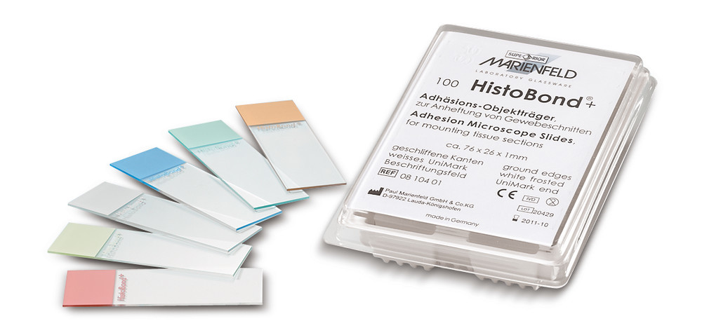
Adhesion slides Histobond ® Corners grounded 90°, natural / AC100034774
MF Part:
CEX0.1
MOQ:
1
£ 34.83
In Stock
more details
Multiple Variants

Adhesion slides Histobond ® Corners grounded 90°, orange / AC100027407
MF Part:
PC71.1
MOQ:
1
£ 37.85
In Stock
more details
Multiple Variants

Adhesion slides Histobond ® Corners grounded 90°, green / AC100021484
MF Part:
PC68.1
MOQ:
1
£ 37.85
In Stock
more details