Products Found:
19
Showing Results For:
Substrates for Biochemistry
Substrates for Biochemistry
Multiple Variants
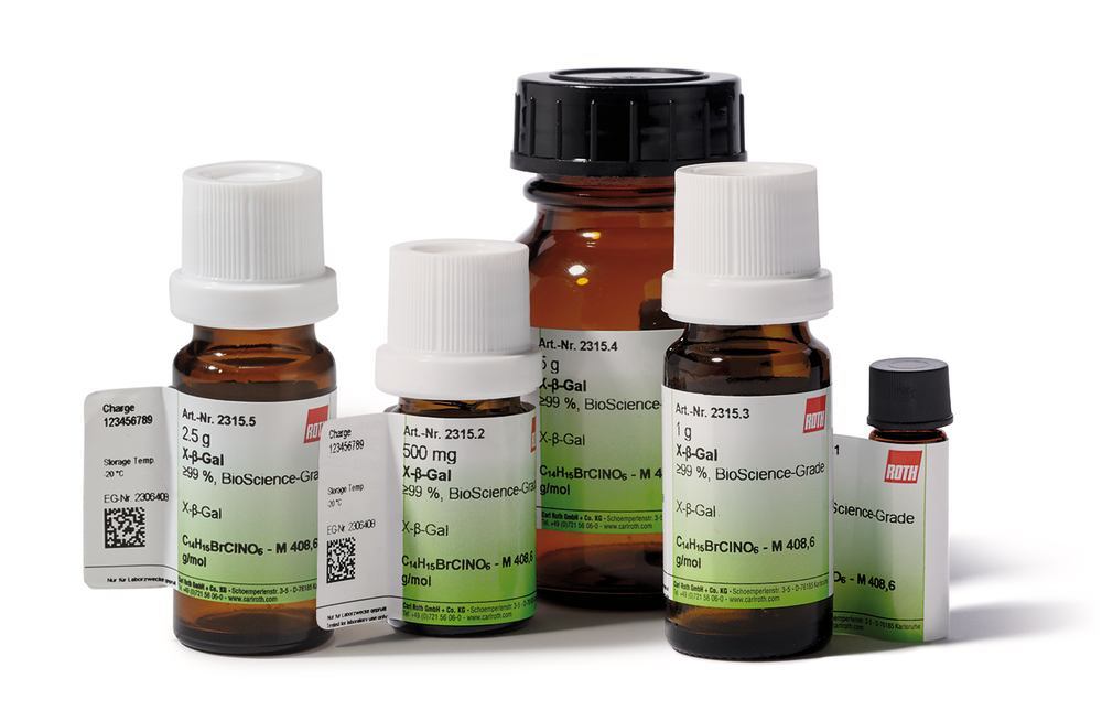
Multiple Variants

Multiple Variants

Multiple Variants

Multiple Variants

4-Methylumbelliferyl-β-D-glucuronide dihydrate, 250 mg / AC100052894
MF Part:
881005-91-0
MOQ:
1
£ 80.24
In Stock
more details
Multiple Variants
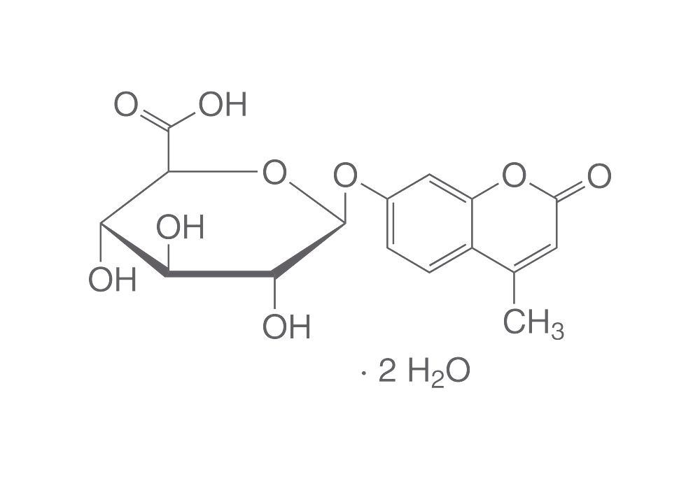
4-Methylumbelliferyl-β-D-glucuronide dihydrate, 50 mg / AC100032382
MF Part:
881005-91-0
MOQ:
1
£ 28.94
Get Quote
more details
Multiple Variants

4-Methylumbelliferyl-β-D-glucuronide dihydrate, 1 g / AC100028721
MF Part:
881005-91-0
MOQ:
1
£ 250.61
In Stock
more details